
Finger Fracture on X-ray & MRI: Imaging Findings
Finger fractures involve the phalanges, the small bones between the knuckles and the fingertip. They can be obvious displaced breaks, tiny avulsion fragments near a tendon or ligament attachment, or subtle injuries that are difficult to separate from a sprain. X-rays are usually the first test because they show alignment, joint involvement, and whether the fracture extends into the growth plate in children. MRI or CT may be used when pain persists despite unclear radiographs or when a clinician needs more detail about the joint surface.
Common Symptoms
- Finger pain, swelling, or bruising after a crush, twist, fall, or ball impact
- Point tenderness over one phalanx or a small painful bump near a joint
- Finger angulation, shortening, overlap, or rotation when making a fist
- Reduced motion because bending or straightening the finger is painful
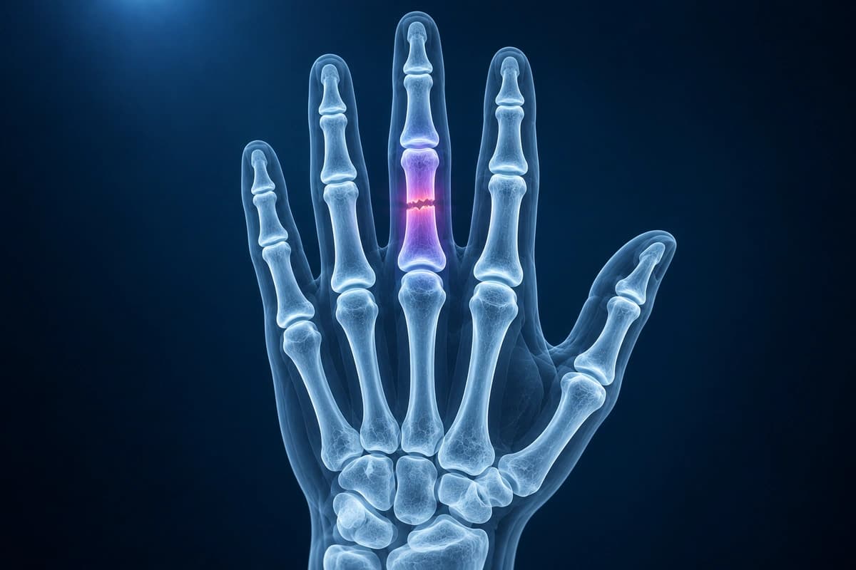
What We Look For on Imaging
- Visible fracture line through the proximal, middle, or distal phalanx
- Small avulsion fragment at a tendon, collateral ligament, or volar plate attachment
- Joint surface step-off, subluxation, or extension into the PIP or DIP joint
- Angulation, shortening, displacement, or malrotation clues on multiple views
- Bone marrow edema on MRI when an occult fracture is suspected
Frequently Asked Questions
Can a finger fracture be missed on the first X-ray?
Yes. Tiny avulsion fragments, nondisplaced fracture lines, and injuries hidden by overlapping fingers can be subtle. Good positioning and at least two views matter. If pain, swelling, or deformity persists despite a reassuring X-ray, clinicians may repeat radiographs or use MRI or CT depending on the suspected injury pattern.
Why does rotation matter in a finger fracture?
A small amount of rotation at the fracture site can make the fingertip cross over its neighbor when making a fist. Imaging can show angulation and displacement, but a clinician also checks finger cascade and motion. That is why X-ray findings should be combined with an in-person hand exam before treatment decisions.
Sources
- Heifner JJ, Rubio F. Fractures of the phalanges. J Hand Surg Eur Vol. 2023;48(2_suppl):18S-26S.
- Oetgen ME, Dodds SD. Non-operative treatment of common finger injuries. Curr Rev Musculoskelet Med. 2008;1(2):97-102.
- Leggit JC, Meko CJ. Acute finger injuries: part II. Fractures, dislocations, and thumb injuries. Am Fam Physician. 2006;73(5):827-834.
- Expert Panel on Musculoskeletal Imaging; Torabi M, Lenchik L, et al. ACR Appropriateness Criteria Acute Hand and Wrist Trauma. J Am Coll Radiol. 2019;16(5S):S7-S17.
- American Academy of Orthopaedic Surgeons. Hand Fractures. OrthoInfo.
Related Articles
Educational red flags for hand and finger infections, including flexor tenosynovitis, abscess, osteomyelitis, and when imaging may be used.
Learn how X-rays and MRI help separate a jammed finger from a fracture, dislocation, volar plate injury, or tendon injury.
Compare finger X-ray, MRI, and ultrasound for fractures, tendon injuries, pulley tears, ligament injuries, and arthritis.
Understand your imaging before your next appointment
MRI, X-ray or CT from a CD, folder or ZIP. A PDF report or photos of the films work too.
Free to open and preview, no account needed. An optional AI report from $10 explains your images in plain language, with questions for your doctor.
Your raw files stay on this device. Only rendered images and minimal context are sent when you request an AI report.
Medical Disclaimer: This page is for informational and educational purposes only. It does not constitute medical advice, diagnosis, or treatment. AI-generated analysis may contain errors. Always consult a qualified healthcare professional for medical decisions. Full Disclaimer