
Metacarpal Fracture on X-ray: Imaging Findings
Metacarpal fractures affect the long bones of the hand between the wrist and knuckles. The fifth metacarpal neck, often called a boxer fracture, is common, but fractures can occur at the base, shaft, neck, or head of any metacarpal. X-rays help determine alignment, angulation, shortening, and whether the joint surface is involved. CT can add detail for complex base fractures or carpometacarpal dislocations.
Common Symptoms
- Pain and swelling over the back of the hand after a punch, fall, or crush injury
- Loss of a normal knuckle contour or a sunken metacarpal head
- Finger overlap, scissoring, or rotation when attempting to make a fist
- Bruising in the palm or tenderness along a metacarpal shaft or base
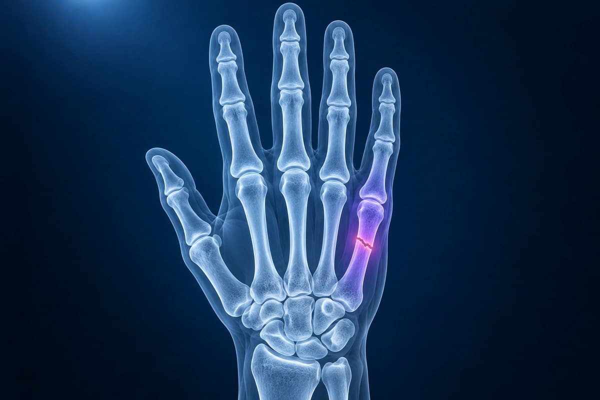
What We Look For on Imaging
- Fracture location at the metacarpal base, shaft, neck, or head
- Volar angulation, shortening, translation, or comminution
- Intra-articular extension into the MCP or carpometacarpal joint
- Associated CMC subluxation or dislocation at the metacarpal base
- Healing callus, delayed union, or hardware position on follow-up imaging
Frequently Asked Questions
Is every boxer fracture treated the same way?
No. Treatment depends on angulation, rotation, shortening, open injury risk, hand dominance, occupation, and which metacarpal is fractured. The little-finger metacarpal tolerates more angulation than the index or middle metacarpal, but rotation is poorly tolerated and usually needs careful clinical assessment.
When is CT useful for a metacarpal fracture?
CT is most useful when the fracture involves a joint surface, the metacarpal base, or a suspected carpometacarpal fracture-dislocation. It can show small fragments and joint step-off more clearly than plain X-ray, which may help surgical planning.
Sources
- Kollitz KM, Hammert WC, Vedder NB, Huang JI. Metacarpal fractures: treatment and complications. Hand (N Y). 2014;9(1):16-23.
- Poolman RW, Goslings JC, Lee JB, et al. Conservative treatment for closed fifth (small finger) metacarpal neck fractures. Cochrane Database Syst Rev. 2005;2005(3):CD003210.
- Fuller JB, Piscoya AS, Clark DM, et al. Surgical management of ulnar metacarpal base fracture-dislocations: a systematic review. Hand (N Y). 2022;17(3):405-411.
- Expert Panel on Musculoskeletal Imaging; Torabi M, Lenchik L, et al. ACR Appropriateness Criteria Acute Hand and Wrist Trauma. J Am Coll Radiol. 2019;16(5S):S7-S17.
Understand your imaging before your next appointment
MRI, X-ray or CT from a CD, folder or ZIP. A PDF report or photos of the films work too.
Free to open and preview, no account needed. An optional AI report from $10 explains your images in plain language, with questions for your doctor.
Your raw files stay on this device. Only rendered images and minimal context are sent when you request an AI report.
Medical Disclaimer: This page is for informational and educational purposes only. It does not constitute medical advice, diagnosis, or treatment. AI-generated analysis may contain errors. Always consult a qualified healthcare professional for medical decisions. Full Disclaimer