
Mallet Finger on X-ray & MRI: Imaging Findings
Mallet finger is an injury to the terminal extensor tendon at the DIP joint. The tendon may tear without a visible fracture, or it may pull off a small piece of bone from the distal phalanx. X-rays are important because a large bony fragment or DIP subluxation can change treatment planning. MRI or ultrasound can show the tendon gap when the X-ray is normal but the fingertip cannot actively extend.
Common Symptoms
- Drooping fingertip after a ball strike, crush injury, or forced DIP flexion
- Inability to actively straighten the DIP joint while passive motion remains possible
- Pain, swelling, or bruising around the back of the fingertip joint
- Small bony fragment near the dorsal base of the distal phalanx on X-ray
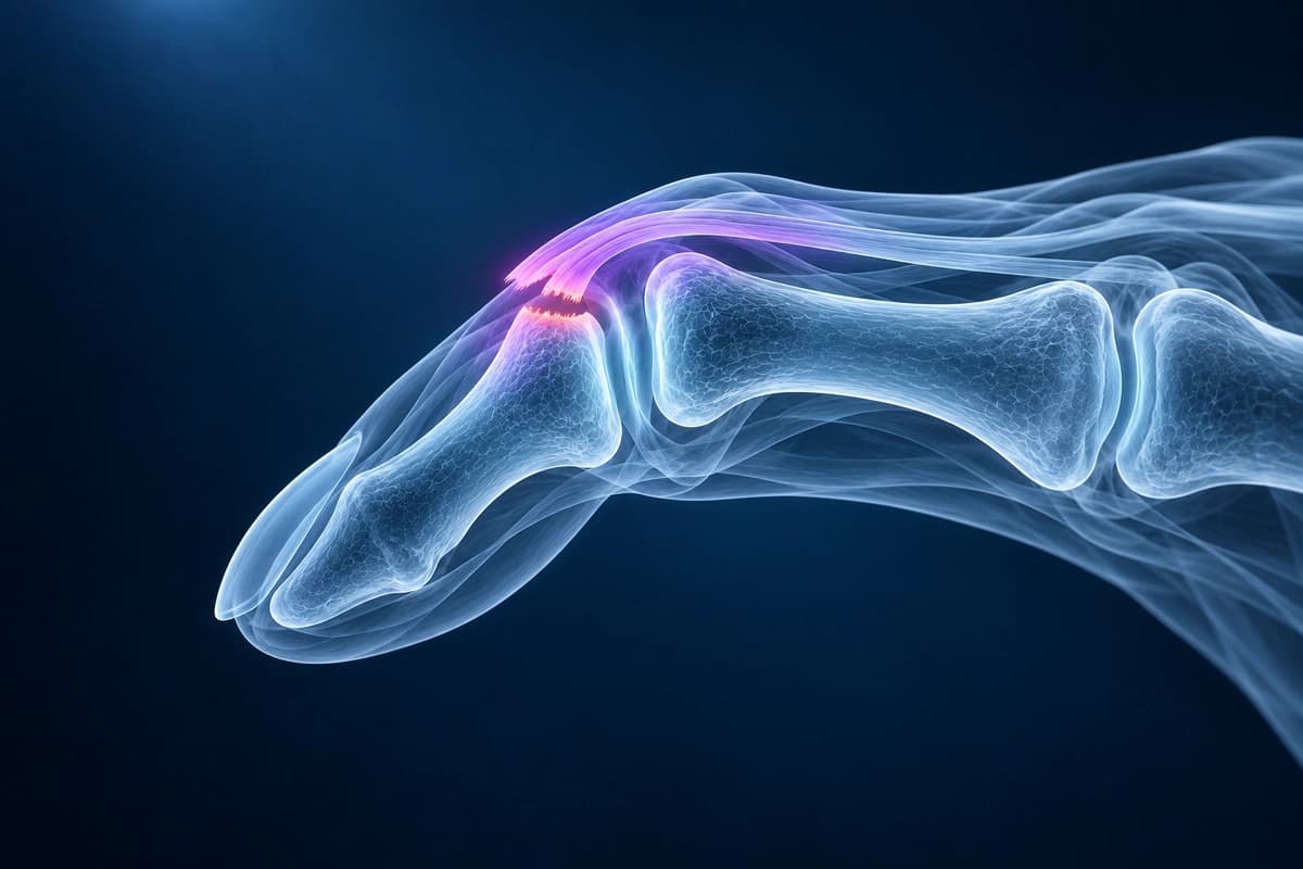
What We Look For on Imaging
- Dorsal avulsion fracture at the base of the distal phalanx
- DIP joint flexion posture or volar subluxation of the distal phalanx
- Size of the articular fragment and percentage of joint surface involved
- Terminal extensor tendon discontinuity, thickening, or retraction on MRI
- Chronic swan-neck posture or degenerative DIP change on delayed imaging
Frequently Asked Questions
Why does a mallet finger usually need an X-ray?
The visible droop can come from a pure tendon tear or a bony avulsion. X-ray helps show whether the joint surface is involved and whether the DIP joint remains aligned. Those details affect splinting strategy and whether a hand specialist should review the injury urgently.
Can AI tell whether a mallet finger needs surgery?
Treatment depends on joint alignment, fracture size, skin condition and examination, with many closed injuries managed by splinting. AI cannot decide whether surgery is needed.
Sources
- Bendre AA, Hartigan BJ, Kalainov DM. Mallet finger. J Am Acad Orthop Surg. 2005;13(5):336-344.
- Salazar Botero S, Hidalgo Diaz JJ, Benaïda A, et al. Review of acute traumatic closed mallet finger injuries in adults. Arch Plast Surg. 2016;43(2):134-144.
- Husain SN, Dietz JF, Kalainov DM, Lautenschlager EP. A biomechanical study of distal interphalangeal joint subluxation after mallet fracture injury. J Hand Surg Am. 2008;33(1):26-30.
- Clavero JA, Alomar X, Monill JM, et al. MR imaging of ligament and tendon injuries of the fingers. Radiographics. 2002;22(2):237-256.
Related Articles
Compare mallet finger and jersey finger injuries, including extensor vs flexor tendon anatomy, X-ray findings, MRI clues, and treatment urgency.
Compare finger X-ray, MRI, and ultrasound for fractures, tendon injuries, pulley tears, ligament injuries, and arthritis.
A plain-language guide to your hand or finger X-ray: how bones and joints should line up, and what fractures, avulsion fragments and arthritis look like.
Understand your imaging before your next appointment
MRI, X-ray or CT from a CD, folder or ZIP. A PDF report or photos of the films work too.
Free to open and preview, no account needed. An optional AI report from $10 explains your images in plain language, with questions for your doctor.
Your raw files stay on this device. Only rendered images and minimal context are sent when you request an AI report.
Medical Disclaimer: This page is for informational and educational purposes only. It does not constitute medical advice, diagnosis, or treatment. AI-generated analysis may contain errors. Always consult a qualified healthcare professional for medical decisions. Full Disclaimer