
Trigger Finger on MRI & X-ray: Imaging Findings
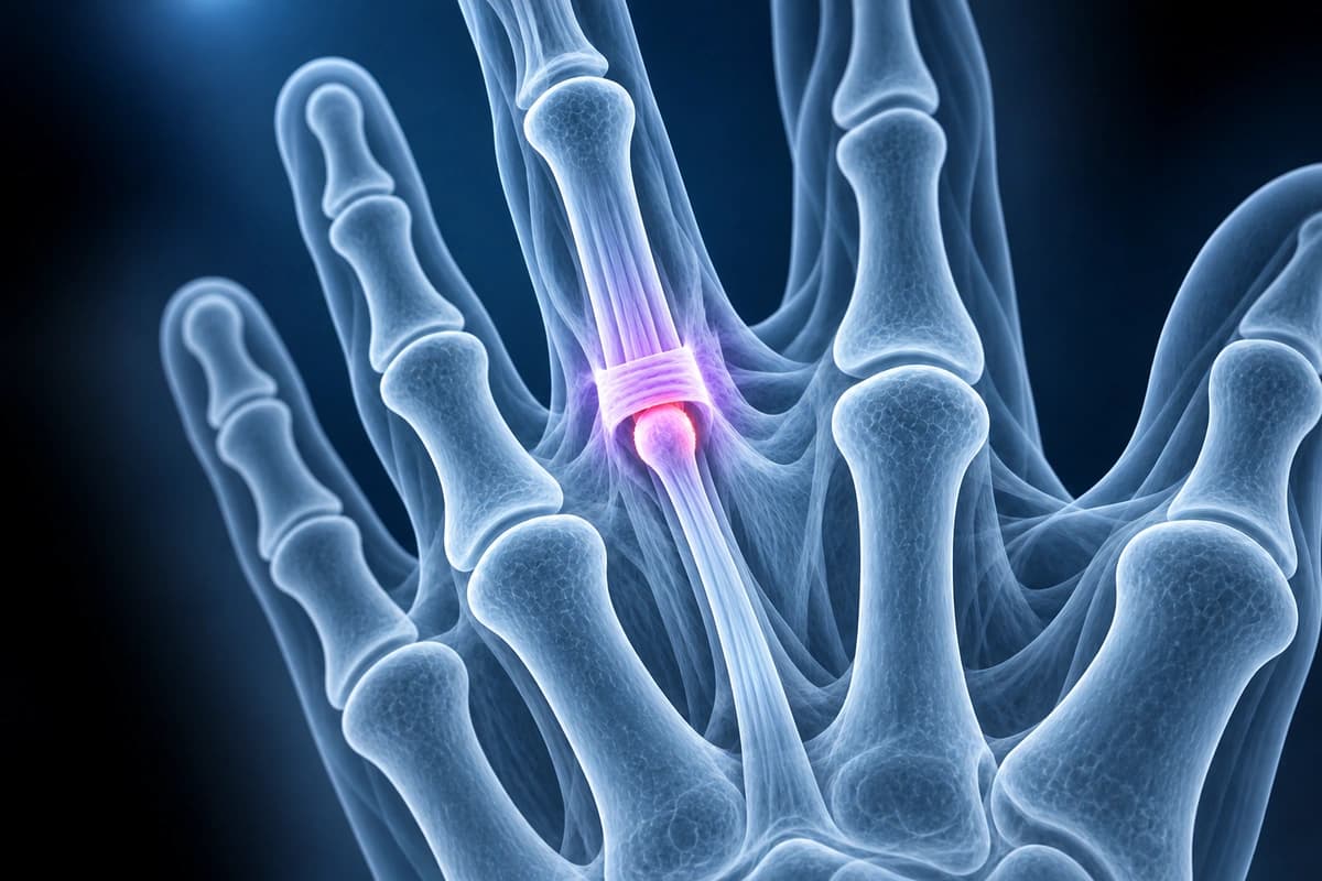
Trigger finger, or stenosing tenosynovitis, occurs when the flexor tendon catches or locks as it glides through the A1 pulley at the base of the finger or thumb. Thickening of the A1 pulley, nodular tendon changes, or tenosynovitis create a size mismatch between the tendon and pulley, causing mechanical catching during finger flexion and extension. The condition is common in individuals with diabetes, rheumatoid arthritis, and those performing repetitive gripping. MRI and ultrasound can visualize pulley thickening, tendon nodules, and associated tenosynovitis.
Common Symptoms
- Catching or clicking sensation when actively bending or straightening the affected finger
- Finger locking in a flexed position that requires passive manipulation to extend
- Pain and tenderness at the base of the affected finger directly over the A1 pulley at the metacarpophalangeal joint
- Palpable nodule in the flexor tendon at the level of the distal palmar crease
- Morning stiffness and locking that partially improves with repeated movement throughout the day
- Multiple simultaneously affected digits, particularly in patients with diabetes or rheumatoid arthritis
What We Look For on Imaging
- Thickening of the A1 pulley beyond normal 0.5 millimeter thickness
- Fusiform nodule or thickening of the flexor tendon at the pulley level
- Fluid within the flexor tendon sheath indicating tenosynovitis
- Loss of normal tendon gliding with dynamic imaging assessment
- Tendon signal abnormality suggesting tendinopathy or partial tear
- Associated findings such as flexor tenosynovitis in rheumatoid arthritis
Frequently Asked Questions
What imaging findings confirm trigger finger and guide treatment planning?
MRI and high-resolution ultrasound demonstrate A1 pulley thickening beyond the normal 0.5 mm threshold, fusiform nodular thickening or increased T2 signal in the flexor digitorum superficialis and profundus tendons at the pulley level, and fluid within the flexor tendon sheath indicating tenosynovitis. Dynamic ultrasound can directly visualize tendon catching at the pulley during active flexion-extension. When trigger finger occurs in the context of rheumatoid arthritis, MRI reveals diffuse flexor tenosynovitis with synovial proliferation and tendon erosion that alters the surgical approach. AI can help explain these imaging terms, but it cannot confirm why the tendon is catching or whether an inflammatory condition is involved. A clinician must assess the cause.
Why is trigger finger more prevalent in diabetic patients and how does diabetes affect treatment response?
Advanced glycation end-products accumulate in collagen-rich structures including the A1 pulley and flexor tendon sheath, causing diffuse thickening and reduced compliance in patients with longstanding diabetes. The lifetime prevalence of trigger finger in diabetic patients reaches 10-20% compared to 2-3% in the general population, and multiple digits are commonly affected simultaneously. Response rates to corticosteroid injection are substantially lower in diabetic patients, approximately 50-60% compared to 80-90% in non-diabetic patients, partly because glucocorticoids transiently elevate blood glucose and partly because the glycation-induced pathology responds less favorably to anti-inflammatory treatment. Earlier consideration of A1 pulley release surgery is therefore appropriate in diabetic patients with persistent triggering.
What is the evidence-based treatment algorithm for trigger finger from conservative to surgical?
Conservative management begins with splinting the metacarpophalangeal joint in extension at 0 to 15 degrees while allowing interphalangeal joint motion, combined with anti-inflammatory medication and activity modification. Corticosteroid injection into the flexor tendon sheath resolves triggering in 60-90% of primary cases, with higher success in non-diabetic patients and early-stage disease. Two to three injections may be offered at six-to-eight-week intervals before surgical referral. Percutaneous A1 pulley release using a needle under ultrasound guidance is an effective office-based alternative. Open A1 pulley release carries a near 100% success rate with minimal recurrence. MRI evidence of extensive tendon degeneration, partial tear, or rheumatoid tenosynovitis supports earlier surgical planning over repeated injection attempts.
Sources
- Makkouk AH, Oetgen ME, Swigart CR, Dodds SD. Trigger finger: etiology, evaluation, and treatment. Curr Rev Musculoskelet Med. 2008;1(2):92-96.
- Guerini H, Pessis E, Theumann N, et al. Sonographic appearance of trigger fingers. J Ultrasound Med. 2008;27(10):1407-1413.
- Fleisch SB, Spindler KP, Lee DH. Corticosteroid injections in the treatment of trigger finger: a level I and II systematic review. J Am Acad Orthop Surg. 2007;15(3):166-171.
- Baumgarten KM, Gerlach D, Boyer MI. Corticosteroid injection in diabetic patients with trigger finger. A prospective, randomized, controlled double-blinded study. J Bone Joint Surg Am. 2007;89(12):2604-2611.
- American Academy of Orthopaedic Surgeons. Trigger Finger. OrthoInfo.
Related Articles
A guide to A2 pulley injuries in climbers, including bowstringing, MRI and ultrasound findings, grading, and return-to-climbing context.
Compare finger X-ray, MRI, and ultrasound for fractures, tendon injuries, pulley tears, ligament injuries, and arthritis.
A plain-language guide to your hand or finger X-ray: how bones and joints should line up, and what fractures, avulsion fragments and arthritis look like.
Understand your imaging before your next appointment
MRI, X-ray or CT from a CD, folder or ZIP. A PDF report or photos of the films work too.
Free to open and preview, no account needed. An optional AI report from $10 explains your images in plain language, with questions for your doctor.
Your raw files stay on this device. Only rendered images and minimal context are sent when you request an AI report.
Medical Disclaimer: This page is for informational and educational purposes only. It does not constitute medical advice, diagnosis, or treatment. AI-generated analysis may contain errors. Always consult a qualified healthcare professional for medical decisions. Full Disclaimer