
Climber's Finger Pain: A2 Pulley Injuries Explained
A guide to A2 pulley injuries in climbers, including bowstringing, MRI and ultrasound findings, grading, and return-to-climbing context.
Key Takeaways
- A2 pulley injuries are common search concerns in climbers
- Ultrasound can show dynamic bowstringing
- MRI can show pulley edema and associated soft-tissue injury
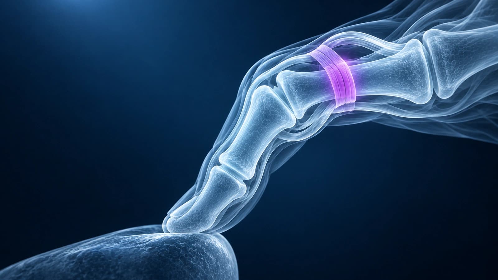
The A2 pulley holds the flexor tendons close to the proximal phalanx. Climbers stress it heavily in crimp grip. A painful pop, palm-side finger pain, or visible bowstringing may lead clinicians to consider a finger pulley injury.
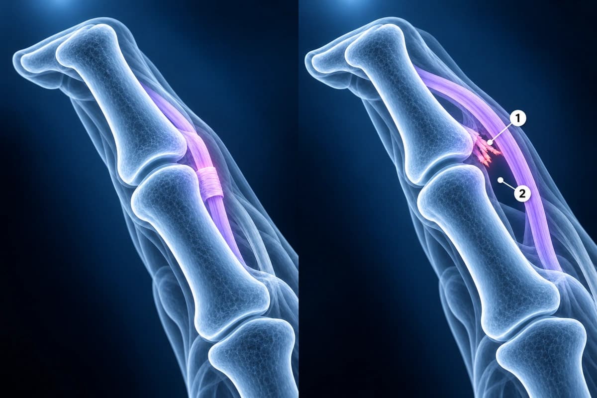
Imaging Signs of Pulley Injury
- Increased tendon-to-bone distance during resisted flexion
- A2 pulley thickening, edema, discontinuity, or nonvisualization
- Flexor tendon sheath fluid or reactive tenosynovitis
- Multiple pulley involvement when bowstringing is pronounced
Frequently Asked Questions
Can I keep climbing with a suspected pulley injury?
Return to climbing depends on grade, pain, bowstringing, strength, and clinician or therapist guidance. Imaging can help characterize the injury but does not replace a supervised return-to-load plan.
Should I pay for 3T MRI for an A2 pulley injury?
Only if it answers a question your clinician cannot answer with exam, X-ray, or dynamic ultrasound. Ultrasound can show bowstringing during motion. MRI, including 3T MRI, can show pulley edema, discontinuity, and associated soft-tissue injury.
What does bowstringing mean?
Bowstringing means the flexor tendon lifts away from the bone because the pulley restraint is torn or stretched. It can be measured dynamically with ultrasound or seen on MRI when the separation is large.
Sources
- Schöffl VR, Schöffl I. Injuries to the finger flexor pulley system in rock climbers: current concepts. J Hand Surg Am. 2006;31(4):647-654.
- Bollen SR. Injury to the A2 pulley in rock climbers. J Hand Surg Br. 1990;15(2):268-270.
- Klauser A, Frauscher F, Bodner G, et al. Finger pulley injuries in extreme rock climbers: depiction with dynamic US. Radiology. 2002;222(3):755-761.
- McCarthy CL. Ultrasound of normal and injured ligaments and retinacula of the hand. Semin Musculoskelet Radiol. 2020;24(2):83-100.
Related Articles
Compare finger X-ray, MRI, and ultrasound for fractures, tendon injuries, pulley tears, ligament injuries, and arthritis.
Learn when 3T hand MRI can help for thumb UCL tears, pulley injuries, occult fractures, and arthritis, and when X-ray, ultrasound, or 1.5T MRI is enough.
A plain-language guide to your hand or finger X-ray: how bones and joints should line up, and what fractures, avulsion fragments and arthritis look like.
Related Conditions
Understand your imaging before your next appointment
MRI, X-ray or CT from a CD, folder or ZIP. A PDF report or photos of the films work too.
Free to open and preview, no account needed. An optional AI report from $10 explains your images in plain language, with questions for your doctor.
Your raw files stay on this device. Only rendered images and minimal context are sent when you request an AI report.
Medical Disclaimer: This page is for informational and educational purposes only. It does not constitute medical advice, diagnosis, or treatment. AI-generated analysis may contain errors. Always consult a qualified healthcare professional for medical decisions. Full Disclaimer