
Hand Arthritis on X-ray: Imaging Findings
Hand arthritis commonly affects the DIP joints near the fingertips, the PIP joints in the middle of the fingers, and the thumb base. X-rays are central because they show joint space narrowing, osteophytes, alignment, erosions, and the distribution of affected joints. MRI is not usually needed for typical osteoarthritis, but it may help when inflammation, infection, occult fracture, or another diagnosis is being considered.
Common Symptoms
- Finger stiffness, aching, or swelling that is worse with use or gripping
- Bony bumps near the DIP, PIP, or thumb base joints
- Reduced pinch strength, thumb base pain, or difficulty opening jars
- Joint enlargement, crepitus, or gradual loss of finger motion
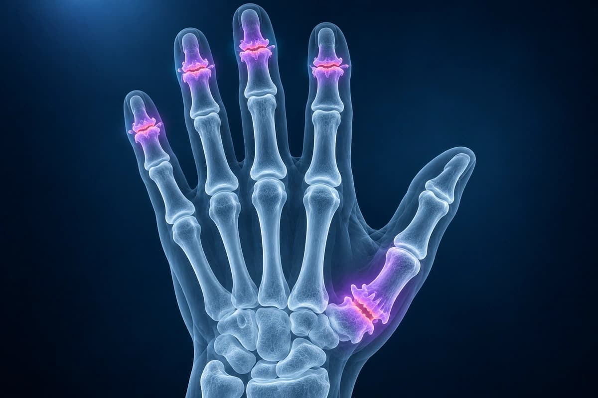
What We Look For on Imaging
- Joint space narrowing at DIP, PIP, MCP, or thumb CMC joints
- Osteophytes, subchondral sclerosis, and cystic change
- Thumb base subluxation or trapeziometacarpal degeneration
- Erosive or inflammatory features that differ from ordinary osteoarthritis
- Alignment changes, deformity, or prior fracture-related arthritis
Frequently Asked Questions
Can an X-ray tell what type of hand arthritis I have?
X-ray patterns can strongly suggest osteoarthritis, inflammatory arthritis, gout, or prior injury-related arthritis, but diagnosis also depends on symptoms, exam, lab tests, and medical history. Imaging is one piece of the clinical picture.
Why does thumb base arthritis matter so much for daily activities?
The thumb CMC joint is heavily used for pinch and grip. Even moderate degeneration can make keys, jars, phones, and writing painful. X-rays help show joint narrowing, subluxation, and osteophytes that may guide bracing, injections, therapy, or surgical discussion.
Sources
- Jacobson JA, Girish G, Jiang Y, Sabb BJ. Radiographic evaluation of arthritis: degenerative joint disease and variations. Radiology. 2008;248(3):737-747.
- Zhang W, Doherty M, Leeb BF, et al. EULAR evidence-based recommendations for the diagnosis of hand osteoarthritis: report of a task force of ESCISIT. Ann Rheum Dis. 2009;68(1):8-17.
- Sakellariou G, Conaghan PG, Zhang W, et al. EULAR recommendations for the use of imaging in the clinical management of peripheral joint osteoarthritis. Ann Rheum Dis. 2017;76(9):1484-1494.
- Kloppenburg M, Kroon FP, Blanco FJ, et al. 2018 update of the EULAR recommendations for the management of hand osteoarthritis. Ann Rheum Dis. 2019;78(1):16-24.
Related Articles
See how osteoarthritis, rheumatoid arthritis and gout look on a hand X-ray: joint space narrowing, bone spurs, erosions and which joints are affected.
Educational red flags for hand and finger infections, including flexor tenosynovitis, abscess, osteomyelitis, and when imaging may be used.
Learn when 3T hand MRI can help for thumb UCL tears, pulley injuries, occult fractures, and arthritis, and when X-ray, ultrasound, or 1.5T MRI is enough.
Understand your imaging before your next appointment
MRI, X-ray or CT from a CD, folder or ZIP. A PDF report or photos of the films work too.
Free to open and preview, no account needed. An optional AI report from $10 explains your images in plain language, with questions for your doctor.
Your raw files stay on this device. Only rendered images and minimal context are sent when you request an AI report.
Medical Disclaimer: This page is for informational and educational purposes only. It does not constitute medical advice, diagnosis, or treatment. AI-generated analysis may contain errors. Always consult a qualified healthcare professional for medical decisions. Full Disclaimer