
Arthritis on Hand X-ray: OA vs Rheumatoid vs Gout Signs
See how osteoarthritis, rheumatoid arthritis and gout look on a hand X-ray: joint space narrowing, bone spurs, erosions and which joints are affected.
Key Takeaways
- Distribution matters as much as severity in hand arthritis
- X-ray patterns suggest diagnoses but do not replace clinical evaluation
- MRI or ultrasound may be used when active inflammation is the question
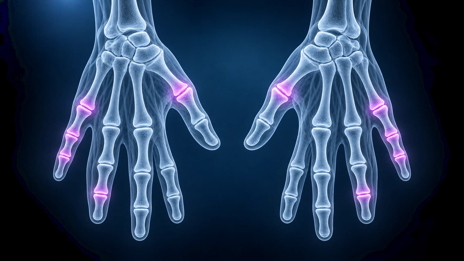
Hand arthritis X-rays are read by pattern. Osteoarthritis often produces osteophytes and DIP or thumb base narrowing. Rheumatoid arthritis often favors MCP and PIP joints with erosions and soft-tissue swelling. Gout can create punched-out erosions and soft-tissue masses.
Common X-ray Pattern Clues
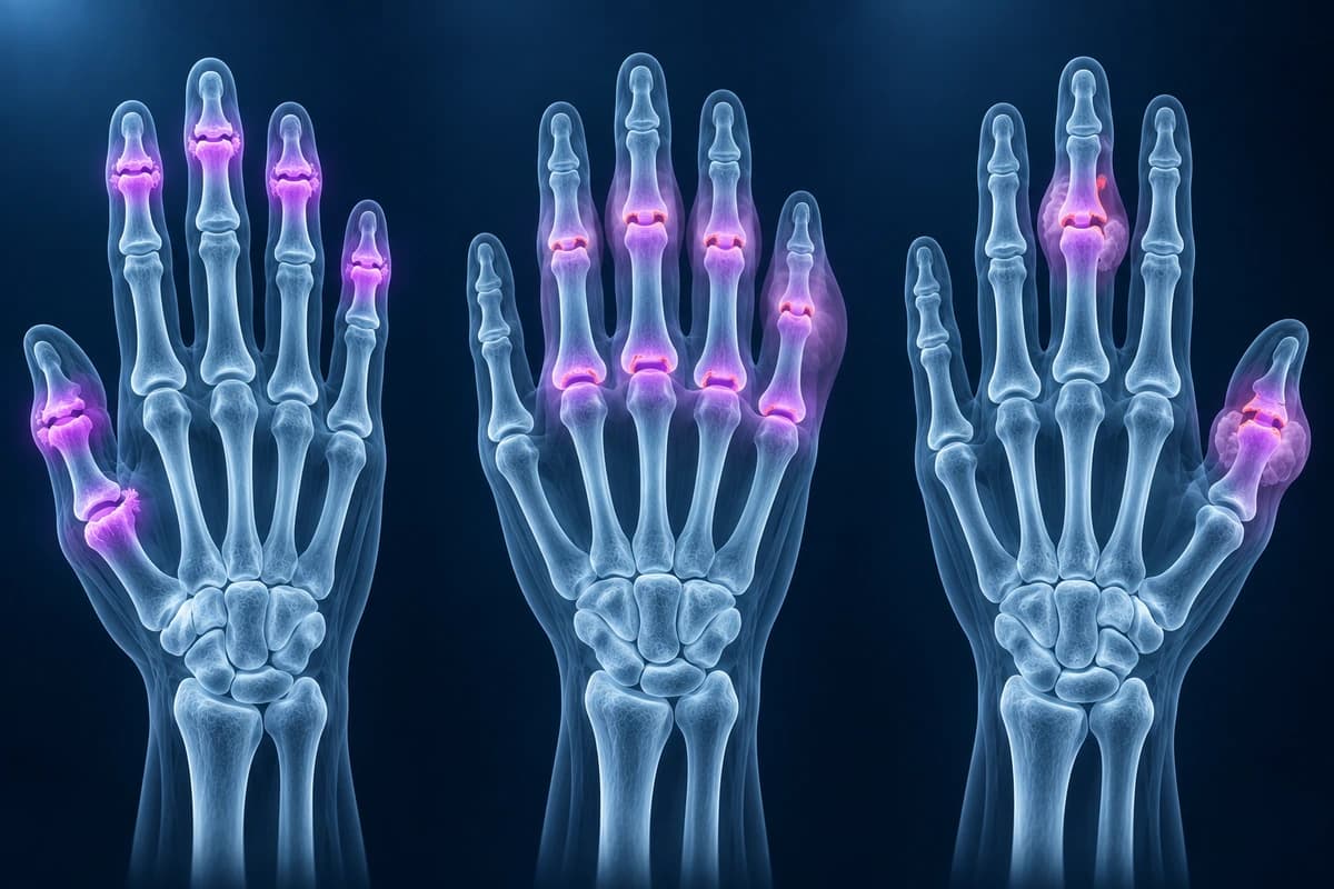
- Osteoarthritis: osteophytes, sclerosis, cysts, and asymmetric narrowing
- Rheumatoid arthritis: marginal erosions, osteopenia, and uniform narrowing
- Gout: overhanging erosions and soft-tissue tophi when advanced
- Post-traumatic arthritis: degeneration centered on an old fracture or joint injury
Frequently Asked Questions
Can X-ray show active inflammation?
X-ray shows structural change better than active inflammation. MRI or ultrasound can show synovitis and tenosynovitis more directly when that question affects treatment.
Should I pay for 3T MRI for hand arthritis?
Usually not for routine osteoarthritis grading. X-ray often shows the distribution and structural severity well. MRI is more relevant when active synovitis, tenosynovitis, early erosions, marrow edema, or an inflammatory arthritis treatment question is being evaluated.
Why do both hands often get imaged for arthritis?
Comparing both hands can reveal symmetry, distribution, erosions, and subtle differences that help separate osteoarthritis, inflammatory arthritis, and other causes.
Sources
- Jacobson JA, Girish G, Jiang Y, Resnick D. Radiographic evaluation of arthritis: inflammatory conditions. Radiology. 2008;248(2):378-389.
- Jacobson JA, Girish G, Jiang Y, Sabb BJ. Radiographic evaluation of arthritis: degenerative joint disease and variations. Radiology. 2008;248(3):737-747.
- Kellgren JH, Lawrence JS. Radiological assessment of osteo-arthrosis. Ann Rheum Dis. 1957;16(4):494-502.
- Eaton RG, Glickel SZ. Trapeziometacarpal osteoarthritis. Staging as a rationale for treatment. Hand Clin. 1987;3(4):455-471.
Related Articles
A plain-language guide to your hand or finger X-ray: how bones and joints should line up, and what fractures, avulsion fragments and arthritis look like.
Learn when 3T hand MRI can help for thumb UCL tears, pulley injuries, occult fractures, and arthritis, and when X-ray, ultrasound, or 1.5T MRI is enough.
Compare finger X-ray, MRI, and ultrasound for fractures, tendon injuries, pulley tears, ligament injuries, and arthritis.
Related Conditions
Understand your imaging before your next appointment
MRI, X-ray or CT from a CD, folder or ZIP. A PDF report or photos of the films work too.
Free to open and preview, no account needed. An optional AI report from $10 explains your images in plain language, with questions for your doctor.
Your raw files stay on this device. Only rendered images and minimal context are sent when you request an AI report.
Medical Disclaimer: This page is for informational and educational purposes only. It does not constitute medical advice, diagnosis, or treatment. AI-generated analysis may contain errors. Always consult a qualified healthcare professional for medical decisions. Full Disclaimer