
Rheumatoid Arthritis in the Hand on X-ray: Imaging Findings
Rheumatoid arthritis in the hand often targets the MCP and PIP joints and can also involve the wrist and flexor or extensor tendons. X-rays look for symmetric joint space narrowing, marginal erosions, soft-tissue swelling, and alignment change. MRI and ultrasound can detect synovitis and erosions earlier than radiographs, but treatment decisions belong with a rheumatology or hand-care team.
Common Symptoms
- Morning stiffness, swelling, or tenderness across multiple MCP and PIP joints
- Symmetric hand pain with reduced grip strength and fatigue
- Ulnar drift, tendon irritation, or changing finger alignment over time
- Known inflammatory arthritis with concern for erosions or active synovitis
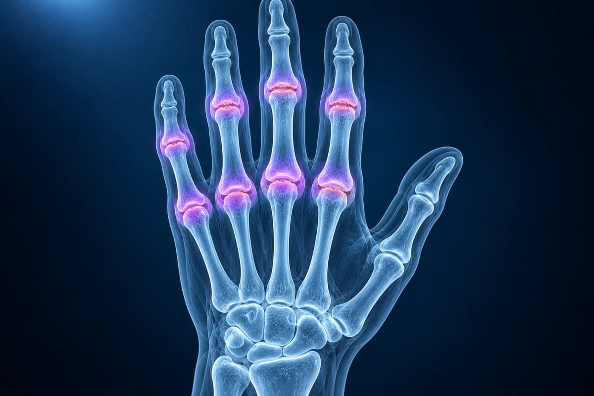
What We Look For on Imaging
- MCP and PIP joint soft-tissue swelling, narrowing, and marginal erosions
- Ulnar deviation, subluxation, or tendon imbalance patterns
- Synovitis, tenosynovitis, and bone marrow edema on MRI
- Wrist involvement that may coexist with finger joint disease
- Secondary osteoarthritis, ankylosis, or prior surgical changes
Frequently Asked Questions
How is rheumatoid arthritis different from hand osteoarthritis on X-ray?
Rheumatoid arthritis often favors MCP and PIP joints with marginal erosions, periarticular osteopenia, soft-tissue swelling, and symmetric narrowing. Osteoarthritis more often shows osteophytes, sclerosis, and DIP or thumb base involvement. Real patients can have overlap, so clinical context matters.
Can MRI show rheumatoid arthritis before X-ray changes appear?
MRI can show synovitis, bone marrow edema, tenosynovitis, and early erosions before they are obvious on radiographs. It is usually ordered when the result will affect diagnosis or treatment planning, not as a replacement for rheumatology assessment.
Sources
- Aletaha D, Neogi T, Silman AJ, et al. 2010 rheumatoid arthritis classification criteria: an American College of Rheumatology/European League Against Rheumatism collaborative initiative. Ann Rheum Dis. 2010;69(9):1580-1588.
- Colebatch AN, Edwards CJ, Østergaard M, et al. EULAR recommendations for the use of imaging of the joints in the clinical management of rheumatoid arthritis. Ann Rheum Dis. 2013;72(6):804-814.
- Østergaard M, Peterfy C, Conaghan P, et al. OMERACT Rheumatoid Arthritis Magnetic Resonance Imaging Studies. Core set of MRI acquisitions, joint pathology definitions, and the OMERACT RA-MRI scoring system. J Rheumatol. 2003;30(6):1385-1386.
- Jacobson JA, Girish G, Jiang Y, Resnick D. Radiographic evaluation of arthritis: inflammatory conditions. Radiology. 2008;248(2):378-389.
Related Articles
See how osteoarthritis, rheumatoid arthritis and gout look on a hand X-ray: joint space narrowing, bone spurs, erosions and which joints are affected.
Educational red flags for hand and finger infections, including flexor tenosynovitis, abscess, osteomyelitis, and when imaging may be used.
Learn when 3T hand MRI can help for thumb UCL tears, pulley injuries, occult fractures, and arthritis, and when X-ray, ultrasound, or 1.5T MRI is enough.
Understand your imaging before your next appointment
MRI, X-ray or CT from a CD, folder or ZIP. A PDF report or photos of the films work too.
Free to open and preview, no account needed. An optional AI report from $10 explains your images in plain language, with questions for your doctor.
Your raw files stay on this device. Only rendered images and minimal context are sent when you request an AI report.
Medical Disclaimer: This page is for informational and educational purposes only. It does not constitute medical advice, diagnosis, or treatment. AI-generated analysis may contain errors. Always consult a qualified healthcare professional for medical decisions. Full Disclaimer