
Boutonniere Deformity on X-ray: Imaging Findings
Boutonniere deformity usually begins with injury to the central slip of the extensor tendon over the PIP joint. When the central slip cannot extend the PIP joint, the lateral bands can shift and the finger may develop PIP flexion with DIP hyperextension. X-rays can show avulsion fragments or joint alignment, while MRI or ultrasound can evaluate the extensor mechanism when the diagnosis is uncertain.
Common Symptoms
- PIP joint droop or inability to fully straighten the middle finger joint
- Swelling and tenderness over the back of the PIP joint after trauma
- Gradual PIP flexion with DIP hyperextension after a missed central slip injury
- History of rheumatoid arthritis or inflammatory tendon imbalance in the hand
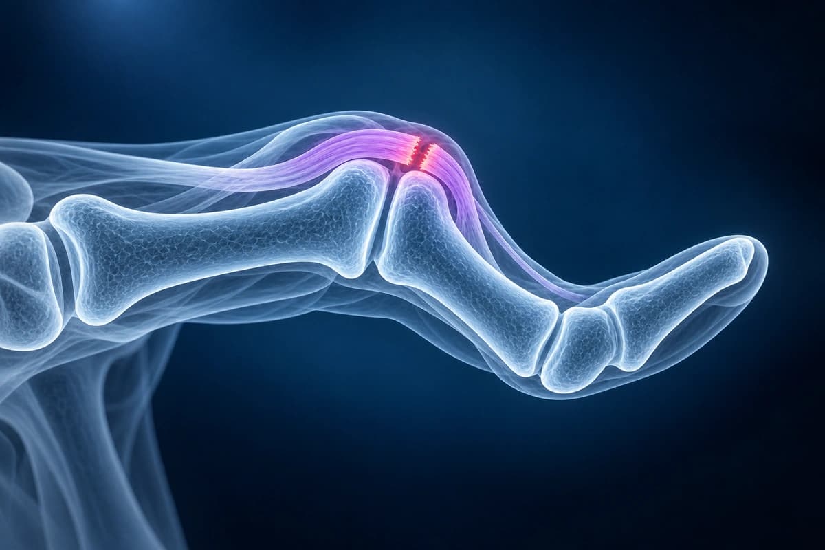
What We Look For on Imaging
- Dorsal avulsion fragment near the base of the middle phalanx
- Central slip discontinuity, thickening, or retraction on MRI or ultrasound
- PIP flexion posture, joint subluxation, or chronic alignment change
- Inflammatory synovitis or erosions when rheumatoid arthritis is suspected
- Associated collateral ligament injury or volar plate scarring
Frequently Asked Questions
Why can boutonniere injuries be missed early?
Early central slip injuries may look like a simple swollen finger, and the fixed deformity can develop later. X-rays may be normal if there is no avulsion fragment. Persistent inability to extend the PIP joint should be assessed clinically even when the radiograph is subtle.
Does imaging replace the central slip exam?
No. Imaging can show avulsion fragments, alignment, and tendon signal, but tests such as active extension assessment and specialist hand examination remain important for deciding splinting, therapy, or surgery.
Sources
- Hanson ZC, Thompson RG, Andrews JR, Lourie GM. Boutonniere versus pseudoboutonniere deformities: pathoanatomy, diagnosis, and treatment. J Hand Surg Am. 2023;48(5):489-497.
- McKeon KE, Lee DH. Posttraumatic boutonnière and swan neck deformities. J Am Acad Orthop Surg. 2015;23(10):623-632.
- Elzinga K, Chung KC. Managing swan neck and boutonniere deformities. Clin Plast Surg. 2019;46(3):329-337.
- Clavero JA, Golanó P, Fariñas O, et al. Extensor mechanism of the fingers: MR imaging-anatomic correlation. Radiographics. 2003;23(3):593-611.
Related Articles
Compare mallet finger and jersey finger injuries, including extensor vs flexor tendon anatomy, X-ray findings, MRI clues, and treatment urgency.
A plain-language guide to your hand or finger X-ray: how bones and joints should line up, and what fractures, avulsion fragments and arthritis look like.
Understand your imaging before your next appointment
MRI, X-ray or CT from a CD, folder or ZIP. A PDF report or photos of the films work too.
Free to open and preview, no account needed. An optional AI report from $10 explains your images in plain language, with questions for your doctor.
Your raw files stay on this device. Only rendered images and minimal context are sent when you request an AI report.
Medical Disclaimer: This page is for informational and educational purposes only. It does not constitute medical advice, diagnosis, or treatment. AI-generated analysis may contain errors. Always consult a qualified healthcare professional for medical decisions. Full Disclaimer